If you want to improve the power of the upper body or prevent injury to excessive use, turn on target forearm exercises in your program is essential. The muscles of the forearm control hand and joint movements and contribute significantly to strength, athletic performance and daily function.
From athletes to office workers, everyone can benefit from stronger forearms. The exercises that target joint muscles and forearms can prevent injuries, improve performance and increase the overall development of hands.
Inspection: What do the forearm muscles do?
Your muscle forearms strength essential movement in the arms, joints and fingers – and to be thoroughly strength. Your forearm muscles support complex movements such as:
Turning the palm up / down
Bending / Enlargement of the wrist and fingers
Move your fingers and thumb in precise grips
Stabilizing the hand while catching or lifting
Are activated in Daily tasks Like wearing groceries, writing, lifting weights, using a computer mouse or playing musical instruments.
KEY CONDITIONS FOR JOINT AND FOREWALS
It does not emphasize due to the storing of each muscle. Just remember:
Flex (front): Bend the wrist and fingers
Extentors (Back): Correct your wrist and fingers
Supinization: Rotate the forearm so that the palm tree turns up.
Workmanship: Rotating the forearm so the palm trees turn downwards.
Radial deviation (Joint kidnapping): tilt the wrist toward the thumb side.
Ulnar deviation (Additeart for the wrist): tilt the wrist towards the small side of the finger.
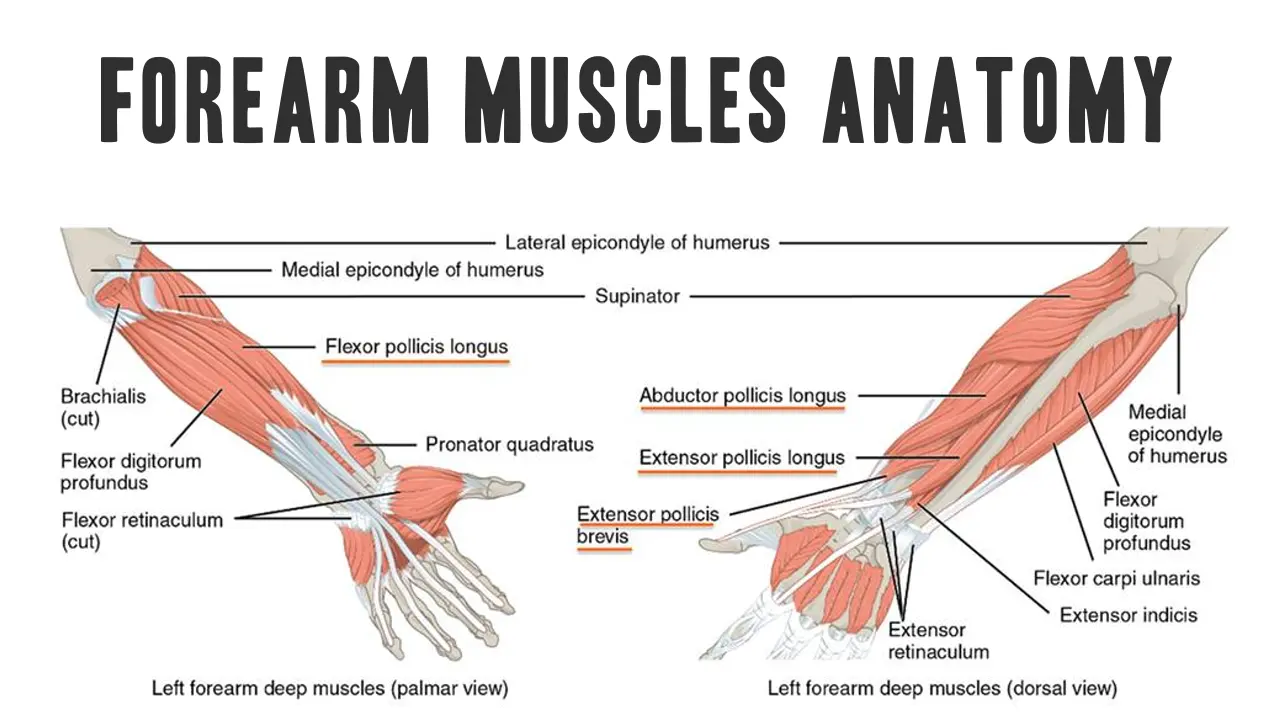
Introduction: Mušići forearm Anatomy
The forearm is the region of the upper limb between the elbow and the wrist. Contains 20 skeletal musclesthat all contribute to the movements of the elbow, wrist and fingers (1). These muscles are organized in Anterior (Flexor-Pronant) and Posterord (Extensor-Supinator) the compartments, each divided into superficial and deep layers.
The compartments of the forearm muscles
1. Front department with anterior (flexor)
Primarily responsible for the flexion of joints and fingers as well as podlaktion. Each section contains superficial and deep layersseparated bones and fibrous membrane.
A-) Surface layer (originates near the elbow):
These are muscles (or structures) that are closest to the surface of the body, just below the skin and subcutaneous fat. Are often the most visible or tangible muscles.
Found Teres: Rotates the forearm inwards.
Carpi Radialis flexor: Flexes the wrist and abducts is (radial deviation).
Palmaris Longus: Helps in the flexion of the wrist.
Carpi Ulnaris flexor: Flexes and adukti wrist.
Flexor digitor: Share in four tendons that help fold the middle fingers and help in the flexible wrist (2).
The Palmaris Longus is absent in about 15% of the population. Its absence does not affect the function of catching or joint significantly (3).
C-) deep layer:
These are muscles (or structure) furthest from the surface, usually lying directly on or very close to the bone.
Flexor Digitorum Deep: Bends the distal finger joints 2-5.
Thumb Longus flexor: Flex the thumb.
Square developments: Rotates the forearm inwards (Pronment).
2 Sentation (Extensor-Supinator) Section
These muscles extend wrist and fingers, help in supinization (Rotation for ignition of roles) and help stabilize the wrist. Originates the most from side epicodile Humerusa.
A-) Surface layer:
Bračionadialis: Folding elbow, helps rotate.
Extensor Digitor Longus and short: Extend and abduct the wrist.
Extension digitor: Prolongs the fingers.
Extension digitor: Extends a small finger.
Extessor Carpi Ulnaris: Extenses and adukti wrist.
Anconeus: Helps expand the forearm in the elbow.
The Extension digitor Share in three sheets: One inserts on Middle Falandand two mergers on Distal Phalanks.
B-) deep layer:
Supinator: Rotates forearm, so Palma turns upwards (Supposition).
Extensor Digitor Longus: Extend your thumb.
Abductor Longus: Moves the palm of the Distance from the palm.
Extensor: Extends the forefinger.
Conditions of ordinary handmade and forearms
The wrist and forearm consisted of a complex bone network, muscle, tendons, ligaments and nerves that allow fine motor control and powerful grip. These structures are vulnerable to excessive use, trauma and postural stress, especially in athletes, workers and manual workers.
Understanding the most common conditions affecting this region is essential for early intervention, appropriate treatment and efficient rehabilitation.
Condition
Cause
Key symptoms
Treatment
Carpal tunnel syndrome
Nerva center compression at the wrist
Stiffness in the thumb / index / medium fingers, night pain
Splinting, nervous slippery, surgery if heavy
Tennis elbow (side epicondilitis)
Provincial of an extensor of the wrist
Pain on external elbow, a weak grip
Vacation, eccentric exercises, forearm
Golferov Lobov (Medial EpiKondilitis)
Propellation of the flexor wrist
Pain on inner elbow, sore throat with joint flexion
Vacation, stretching, eccentric flexing training
Cuervain’s Tenosinovitis
Inflammation APL / EPB tendon
Pain near the thumb base, even worse with the thumb movement
Palac, NSAIDS, Corticosteroid injection
Wrist
A strain of ligaments or tears
Swelling, bruising, limited ROM
Rice, fastening, gradual rehabilitation
Distal radius fractures (colles’)
Falls on the outstretched hand
Deformation handed, pain, swelling
Inflation or surgical fixing
Ulnar Nervous Curability (Guion’s Canal)
Ulnar nerve compression at the joint
Stiffness in pinki / ring fingers, weakness of squeezing
Release the tension and prevent injuries with this:
Wrist rotations: Make fists, rotate your wrists in both directions.
Flexor Flexibility forearms: Hand straight, palm, drag gently.
Extensor forearms are stretched: Hand straight, palm down, pull your hand gently.
The strengthening and stretching of the forearm can promote better function and help prevent injuries.
Don’t ignore them in your exercises.
Train both flexors and extensions.
Treat injuries early.
Keep them with mobile and resistance with prostheses.
Conclusion
The forearm is a complex range composed of 20 muscles that allow precise and powerful wrist movements, arms and digits. Understanding the layer of anatomy, functions and variation is essential for health workers, fitness experts and any interested in human biomechanics. Recognizing common anatomical variants and clinical implications increases diagnostic and therapeutic accuracy.
References
Brittney Mitchell, Lacey is Whit. Anatomy, shoulder and upper limb, forearm muscles
Lauren Okafor, Matthew A. Varaacallo. Anatomy, shoulder and upper limb, hand-flexible digitorum superphizicis
Prevalence of the Palmaris Longus muscle and its relationship with solid strength and strength: study in Turkish pediatric population
Moore KL, Dallei AF, Agur am. Clinically oriented anatomy. 7. ed. LippinCott Williams & Wilkins.
Standard S, ed. Gray’s anatomy: Anatomical basis of clinical practice. 41. Ed.
Saladin Ks. Anatomy and physiology: unity of forms and functions. 9. ed.
Netter FH. Atlas of human anatomy. 7. ed.
Jacobson MD, Raab R, Fazeli BM et al. Anatomical variations of the forearm muscles and their clinical importance. Klin Anat. 2001
ROI J. Variants of Palmaris Longus muscle. J Hand Surg. 1995
Sunderland S. Nerves and injury to nerves. Vol 1 and 2. 1978.
Hirasawa I et al. Studies on the distribution of the superficial branch of the radial nerve. J Hand Surg Am. 1987
Thompson NV, Mockford BJ, Cran GV. The absence of Palmaris Longus muscle. Ulster Med J. 2001
ROI TS et al. Variation in the origin of flexory Pollicis Longus. Klin Anat. 2002
Oh Sj, etc. Anastomosis of Martin and Grude in humans. Muscle nerve. 1976